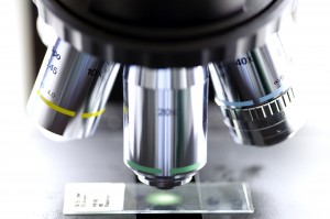
 Darkfield microscopy presumes that the property of your blood can give information about disturbances in the organism.
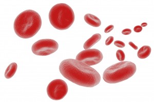
Live cell analysis? Never heard of it? It has been rumoured to give amazing insight. Particularly when you are trying to find an explanation for complaints that are hard to understand. Or, to attest to your therapy’s success, and to detect trouble while it’s still in the early stages, before it could do damage.
When doctors use the term “blood count”, they are referring to the counting of different blood components. These are also called concentrations. But the information Practitioners of Alternative Medicine seek to extract from a drop of blood using Darkfield Microscopy, is of a completely different nature. So what is it they are looking to find under the microscope?
First and foremost, it is obvious that blood is alive. The blood picture in the darkfield is somewhat similar to a photograph taken under water. With the difference that instead of finding fish, jellyfish or star fish, we can see components of blood in different sizes and shapes. It is like seeing a snapshot – as if you were looking through a peephole into an aquarium. It is possible that at the next moment, or when using a different drop of blood, the picture could be different.
In modern clinics, such as K-W Homeopathic Medicine, the patient can often watch their live cell examination because the view into the darkfield microscope is projected onto a screen. Red and white blood cells will appear: at first fresh and mobile. Then later, while the drop of blood dries out, they decompose. Countless other blood particles can become visible, but not necessarily.
Darkfield microscopists assume that blood is not sterile, but houses a number of tiny living organisms. These can include completely harmless little guests (“symbionts”), or other organisms that rob their involuntary host of a good deal of vitality.
Professor Enderlein’s hypothesis
Every properly equipped microscope will make these pictures visible. But in Darkfield Microscopy, it is particularly the interpretation of these signs that forms a part of the technique. In doing so, many therapists are acting on the authority of the German microbiologist and zoologist Prof. Dr. Günther Enderlein. He developed a comprehensive theoretical structure in the early 1900’s, but it is not recognized by mainstream medicine.
Enderlein was certain that even the tiniest protein parts that are visible in the darkfield, could develop into bacteria, viruses or parasites. He named these smallest parts “protits”, after the Greek God Protheus, who was able to transform himself into any shape or form he desired. Thus, for Enderlein and his supporters, viruses, bacteria or fungi are different forms of development of a tiny basic form of life.
But can harmless microbes really change into deadly pathogens because their breeding ground allows it? Every scientist would dispute this. However, the story of Tutankhamun and “The Golden Pharao“ could speak for it.
Some practitioners specializing in Darkfield Microscopy don’t believe in Enderlein’s Theory. They explain away the particles as oxidative stress attacking the cells, thereby causing the cells’ constituents to oxidize. Then once they are released, they combine with blood proteins and show up as “darkfield corpuscles”. Yet, regardless of Enderlein’s interpretation, Darkfield Microscopy has its value in identifying changes in the darkfield blood picture that could indicate a shift in the internal milieu which should be treated.
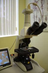
Blood is observed through a darkfield microscope. But the picture can also be transmitted to a screen so that both practitioner and client can observe the image
What does Enderlein’s Theory have to do with
The curse of King Tut’s Tomb?
In 1922, the almost untouched tomb of Tutanchamun was discovered in the Valley of the Kings. An inscription on the tomb read: ”Death will come swiftly to those who disturb the tomb of the king”. But archaeologists opened the tomb which had been closed for thousands of years, anyways. They found the famous golden mask and other treasures.
However, they did not get to enjoy their fame for this discovery for long. Some of them died in the following years under more or less mysterious circumstances, that were thought to be linked to the curse of the pharaoh.
51 years later, twelve out of 14 Polish scientists met with a similar fate: They climbed down into the royal crypt of Krakow in 1973, and died soon thereafter. Yet the Polish, refusing to believe in a curse, had some researchers address the question instead of whether or not fungi such as Aspergillus flavus or Aspergillus niger could be the reason for the deaths. Only: How was it possible for this fungus to develop in a closed tomb?
Some Darkfield Microscopists see Enderlein’s theory confirmed here, in that bacteria can develop into fungi. They believe that Tutanchanum died of tuberculosis and that the tubercle bacteria are an early form of the fungi Aspergillus. In opening the tomb, the archaeologists would have breathed in the deadly fungi that had grown on the thousands-of-years-old corpse.
This gives rise to lots of speculation. The only thing that’s for certain at this point is that modern archaeologists would rather wear face masks when dealing with mummies.
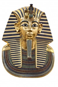
Some researchers explain The curse of King Tut’s Tomb by the fact that obviously, fatally acting mold spores managed to survive in the tombs
How Darkfield Microscopy works
To really understand nature of Darkfield Microscopy, it is best to first imagine the opposite: Brightfield microscopy: Here, some blood is spread onto a glass microscope slide which is then left to dry. In order to recognize the individual blood cells under the microscope, they need to be dyed. You are looking directly into the light source through the magnifiers of the microscope. This is just as difficult as would be the attempt to observe blossoms against the sun.
In the darkfield procedure on the other hand, a drop of blood is transferred onto the slide within seconds and protected with a slide cover, to prevent it from being altered when exposed to air. Because when it dries up, abnormalities can emerge even in healthy blood, which are best avoided as much as possible. Under the microscope, the drop of bood is illuminated via a cone of light only. This way, the blood particles shine like blossoms in a dark box when they are spotlighted from the side.
The most important Signs
A typical sign known to every darkfield practitioner is the phenomenon of the rouleau, in which the red blood cells are stacked one on top the other like coins. What we have here is an agglutination of red blood cells. They are not separated any more as they normally would be, but stick together like stacked coins.
One of the functions of red blood cells is to transport oxygen. In this case, because of the agglutination, their surface is reduced which in turn impairs their ability to carry through the transportation. In addition, the blood becomes thicker. When Natural Health Practitioners see this sign, they assume that a person’s circulation is affected, and that in the case of very fine blood vessels which are damaged already, there may be a potential for “mini infarcts”.
Stacked coins are always a sign of too much acidity in the blood. Our blood normally has a pH value of between 7.35 and 7.45. An acid milieu is found only in the stomach (very acidy: 1.5 – 4!) and in the vagina (4 – 4.5) Everywhere else in the body values under 7.45 are not good. The body does not tolerate even smallest deviations and will react with changes that are visible in the darkfield.
When the blood is too acidy, symbionts can develop further into parasitic forms. Friendly symbionts generally float around in the background of the darkfield like snow flurries without harming their host. They even satisfy regulatory functions, normalizing to some degree disorders which have occurred. But these friends, as mentioned previously, can also turn into enemies. When the terrain becomes overacid on a chronic basis, even if it is only a slight change.
Typical Changes seen in the Darkfield
Stacked Coins:
Overacidity, dehydration, infections
Deformation of red blood cells:
Lack of hemoglobin or vitamin B12, liver stress
Number and motility of white blood cells:
Condition of the immune system, for example infections or allergies
Filits:
Indication of increased oxidative stress (“overacidity”). Filits can lead to poor circulation.
How Cell phone radiation can affect blood
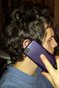
Cell phone usage can lead to stacked coin formation in the blood. High school graduates in Germany made this discovery,
winning them the 1st prize at the “Youth Research”contest.
The connection between the stacked coin phenomenon and cell phone radiation was researched a few years ago. In 2005, two high school grads received the first prize for biology within the “Youth Research” contest. They proved that calls placed on a cell phone can lead to the formation of stacked coins. 51 test persons had one drop of blood taken from then. The differences in the darkfield pictures were striking. The stacked coins were still present after a third drop of blood had been taken– 10 minutes later.
It may be somewhat comforting to know that such changes in the blood will likely disappear in healthy people. After all, the test persons did not use a cell phone for the first time in their lives and their blood did not have stacked coins prior to the phone call. But the thought of our red blood cells being so susceptible to cell phone radiation it isn’t exactly reassuring. So what is happening here?
One of the many possible causes for over-acidity could be electric smog generated during cell phone usage. Some people working in electronics stores show substantial agglutination in the blood. But also an unhealthy diet, lack of sleep, kidney dysfunctions or stress will cause the blood to be acidy and give a stacked coin picture.
Even using the sauna can lead to stacked coin formations. So apart from its therapeutic benefits, a sauna session can obviously irritate the body to a certain degree too. But thanks to the regulating symbionts, stacked coins disappear in healthy people the day after the sauna.
Another reason for blood cells sticking together and thereby prompting problems with circulation could be dehydration, or an acute infection with a large amount of antibodies. The stacked coins seen after using the sauna could possibly also be linked to the heavy sweating.
Darkfield Microscopy in Clinical Application

At K-W Homeopathic Medicine, we use Darkfield Microscopy not to diagnose, but to educate and motivate you and show you the response to nutritional changes, detoxification and life style changes.
While Darkfield Microscopy is used at our clinic as an educational tool to treat a person’s terrain, it also has great value in helping with case assessment. Here are some examples from the practice of fellow practitioners in Germany:
Fit again for work after suffering from chronic fatigue
In someone presenting with chronic fatigue, their blood picture in the darkfield would likely show the person’s cells clumping together. The cells’ job is to transport oxygen, but this function is impaired when a picture like this emerges. Each cell in the body has mitochondria, which like natural mini power plants transform the oxygen from the blood into energy. The better they work, the more productive we feel. But as we age, it is normal that not all mitochondria will work to full capacity. We experience a lack of energy and chronic illnesses can develop.
This case is of a 62-year old who suffered increasingly from exhaustion. He was markedly stressed – both in his career as well as in his private life. The patient underwent prostate surgery in late 2011 but suffered complications, so that the surgery ended up taking 10 hours – all while he was under general anaesthetic!
At the end of his hospital stay, his debility had worsened to the point where he became unable to work. In the darkfield picture of this person, massive agglutinations of red blood cells were seen as well as indications for degenerative changes in the blood components.
He was then started on certain minerals, vitamins and amino acids and at the same time, treated homeopathically using Sanum Therapy, in this case Sanuvis and Sankombi.
Six weeks later another drop of blood was examined under the darkfield microscope. The agglutination of his red blood cells was lesser and so were the degenerative changes in the blood picture. Today, this patient feels well and he has been able to resume work after being on disability for several months.
Observing the changes in one’s darkfield picture is especially important and in this case it had a motivating effect on the patient: It is a visual feedback mechanism about the therapeutic success – completely non-judgmental and without finger wagging.
Finding the right therapy with the help of Darkfield Microscopy
Then there is the case of a 50-year-old lady, a married engineer, who was tired and overworked for years. Years ago, she and her husband tried to conceive unsuccessfully. Fertility treatments including artificial insemination did not work. Although she got pregnant twice, she lost the child within the first weeks of pregnancy. To her it felt as if the baby had “dried up” inside of her. And so they came to terms with having to live a life without kids.
And then at age 50 came the shocking diagnosis: Breast cancer. Shattered, she followed doctors’ recommendations to have the affected breast removed. Afterwards, the initial recommendation was for her to get six chemotherapy treatments. It was during this time that the patient sought help from a natural health practitioner for the first time, as she was increasingly deteriorating. She could no longer work, had lost weight and all of her hair, and felt so sick after each round of chemotherapy that she couldn’t eat or drink.
Her darkfield picture looked horrific. She was chronically overacid, had liver and kidney dysfunctions and her body’s innate ability to self-regulate was hardly in existence. The Natural Health Practitioner initially prescribed vitamins, minerals and homeopathic remedies to make up for the loss of fluids. To supply the body with substances which it had likely been missing for years. But also to normalize organ functions so that she was better able to tolerate the chemotherapy.
Her Natural Health Practitioner recommended she change her diet (oil-protein diet according to Dr. Johanna Budwig). In addition, she was treated isopathically and received herbal medication, as well as the above mentioned protocol. The darkfield live blood picture looked much better two months later. And the patient also felt better. She was also treated with healing hypnosis to direct her thinking into a more positive direction and bring back her enjoyment of life.
Today, four years after her surgery, the patient feels great. She sees her cancer as a process that had signalled its coming for years or even decades: by exhaustion, lack of energy, constant mental overload and the loss of all enjoyment of life. However, she refused to listen to the signals. This patient now comes in regularly for live blood cell analyses.
Ridded of psoriasis
In yet another case, a little boy was diagnosed with psoriasis at the age of 2. Medical and natural treatments were only short-acting or did not give any relief whatsoever. The child scratched himself almost over the whole body, the skin was dry, flaky and red, some areas were bloody or infected.
The first darkfield live blood cell picture showed liver dysfunction and colonization in the colon. A chronic acidity and bacterial stress could also be observed.
Based on her darkfield live blood cell analysis, the German Natural Health Practitioner made some treatment recommendations. She used Notakehl D3 cream, Sanuvis D1 cream as well as ozonated olive oil in order to keep the bacterial infections and the itching at bay.
However, it was more difficult and time-consuming to treat the causes. The child’s diet needed to be adjusted: at first, no animal proteins, no milk and milk products, no sugar and particularly no “bad” fats (like margarine, no chips), using virgin olive oil instead. To build up the child’s intestinal flora, the little patient was given a childrens’ probiotic preparation, as well as a product containing milk thistle to improve liver function.
Initial improvements set in quickly. The child’s complexion and wellbeing improved dramatically, likewise his darkfield live cell picture. But there were small setbacks all the time. When checking back with the family, it turned out that not all members adhered to the dietary instructions, giving the boy chips and chocolate on occasion. The reaction was seen immediately after consumption. But this problem was too eventually solved.
In the meantime, at almost three years old, the little boy is a happy and healthy child. His psoriasis is gone and along with it the agonizing itch. He no longer needs medication. He is also on a well-balanced full-fledged diet. Now he is able to tolerate animal protein well. But his mother was asked to make sure that in future too her son’s diet not include any denatured substances. For example, no white sugar, no sweetened ready-made teas, no homogenized milk.
What are the limitations of this method?
Darkfield microscopy shows a picture of live blood – no more and no less. Its value for Natural Health Practitioners lies in its capacity to make visible subtle changes before pathological changes can be detected by an ordinary lab. It thus allows the practitioner to be proactive by treating a person’s terrain. It is not possible to make a precise diagnosis using darkfield analysis, because it only brings into view any deviations from the normal. The therapist’s knowledge and skill are quite important when it comes to interpreting the darkfield picture.
While the diagnosis of a heart attack is established by an ECG and corresponding lab tests, a diagnosis of cancer is made histologically. Still, darkfield analysis is indispensable for many Natural Health Practitioners. In particular because this method may show abnormalities which appear in the blood picture long before the onset of an illness. It is our aim to ensure by means of appropriate therapy, in treating the person’s milieu, that complaints do not develop into serious problems in the first place. The method also helps those who don’t feel taken seriously because nothing shows up in their lab tests and examinations. Darkfield Microscopy can often help these people by indicating which is the right therapy for them.
New at K-W Homeopathic Medicine and Wellness Clinic
We are very excited to offer Darkfield Microscopy as a new service at K-W Homeopathic Medicine and Wellness Clinic.
If you are coming in for treatment, and if on a scale of one to ten, with ten being the most serious, your condition is an eight, then as you start to implement the recommended protocols, this technology will let you see the changes that occur in your blood as it moves from being a swamp to being a river.
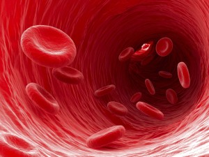
And your blood is a river.
If you clean up the river, you put fresh water everywhere – and all of the nutrients and oxygen that are carried in it.
That’s the real benefit of looking at your live cells, because you can literally see this cleanup process happen.
Introductory Special!
For a limited time, until June 30, 2014, we are offering live cell analysis sessions at
$ 50.00 + HST single session
$ 150.00 + HST package of four sessions
Effective July 1, 2014, prices will be $ 95.00 for a single session, resp. $ 285.00 for a package of four.
Be sure to book your appointment now to take advantage of this offer. Call K-W Homeopathic Medicine and Wellness Clinic at (519) 603-0505 and be among the first to receive this service!
If you have any questions or comments with respect to this article, homeopathy or any other service offered at K-W Homeopathic Medicine and Wellness Clinic, please contact me directly at (519) 603-0505, or speak to one of our practitioners and we will be happy to help!
In health,
Irene Schwens, DHMHS, C. Tran.
Owner / Homeopath,
training and expertise in
Classical Homeopathy and Live Cell Microscopy
Reference:
Bio 2013/1
